Category: Anatomy
The pancreas can be one of the more difficult structures for beginning anatomy students. This is partly because it is a gland that doesn’t preserve well and can fall apart easily when handling the stomach or other organs. In ideal specimens, the pancreas can be seen as a bumpy …
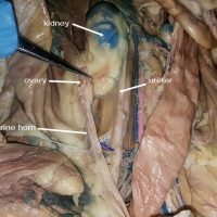
In cats and many animals that have multiple embryos, the uterus is divided into two sides called the uterine horns. The ovary can be found at the end of each horn. In humans, this structure is homologous to the Fallopian Tubes. The photo below shows only the right …
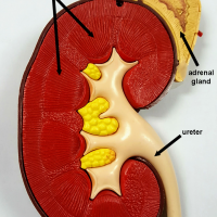
Renal pyramids are triangular shaped areas seen on a cross section of the kidney. They appear striped due to the thousands of nephrons within them that make up the functional unit of the kidney. The nephrons perform the function of filtration of waste products from the blood and …
Students learn the names of bones and their structures for Anatomy and Physiology. This is a much more in-depth study than just labeling the skeleton bones because they also need to know the features of the bones where muscles attach and how the bones fit together. I use …
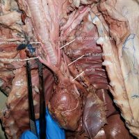
The carotid arteries supply blood to the head and neck. In humans, the right common carotid branches from the brachiocephalic artery and the left common carotid branches directly from the arch of the aorta. On the cat, as shown in the photograph, both common carotids branch from the …
The chordae tendinae are tiny tendons that connect the valves of the heart to the papillary muscle. They appear as small white strings and are sometimes referred to as the “heart strings.” In the photo below the right ventricle has been cut and the chordae shown are attached to …
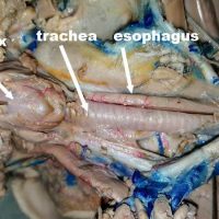
The larynx is also known as the voice box and it can easily be found in the throat of mammal specimens (fetal pig shown below.) The larynx contains the vocal cords that are used for making sounds. In humans the location of the larynx allows for diverse sounds …
The bicuspid and tricuspid valves are both located between the atrium and the ventricle. They are similar in appearance and can be difficult for beginning anatomists to identify accurately. There are a couple of different ways to remember and then locate the valves. First, you must remember …
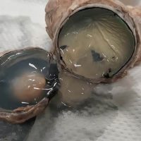
This dissected eye shows the clear gel that fill the space between the lens and the retina of the eye. In a preserved specimen, the fluid is cloudy and thick, having the consistency of gelatin. When the eye is cut in half, the lens can be found floating …
Students in biology are often tasked with dissecting a frog, but there are many other vertebrate models that can provide a rich experience in learning anatomy. Common specimens include the fetal pig, shark, mink, cat, and the rat. Rats are fairly inexpensive and are raised for …
During the cardiac cycle, two contractions occur. Systole occurs when the muscles contract and diastole occurs when the muscles relax. Atrial systole occurs when the atria contract and push blood into the lower ventricles. During this point, the aortic valve is closed. Ventricular contraction then occurs …
The cerebellum is located at the posterior end of the brain and is associated with motor control, balance and coordination. It is only loosely attached to the cerebrum, and dissections of the brain reveal that removing the dura mater often result in the accidental removal of the cerebellum. …
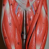
The thigh muscles are often collectively called the quadriceps, which consist of four separate muscles: rectus femoris, vastus lateralis, vastus medialis, and the vastus intermedius. On most diagrams, the rectus femoris is shown as the front muscle that runs parallel to the femur. The word “rectus” means …
The lower arm consists of two bones, the radius and the ulna. The ulna articulates with the humerus to form a hinge joint. The radius articulates with the ulna at the radial notch and forms a pivot joint, allowing for the rotation of the wrist. This type of …
The ulna is one of two bones found in the forearm. It forms a hinge joint with the humerus and a pivot joint with the radius, giving the lower arm the range of motion to bend and rotate the hands. On anatomical drawings, the ulna and radius …
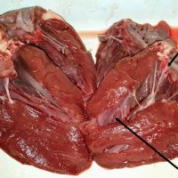
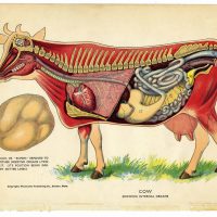
You may have heard that cows have 4 stomachs, but that is not actually accurate. Cows, like all other vertebrates, have the same basic blueprint for the digestive system: mouth –> esophagus –> stomach –> small intestine –> large intestine –> anus There is one stomach, but that stomach is …
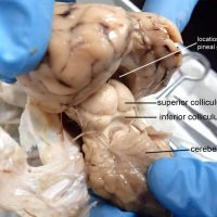
The superior colliculus is part of the midbrain, also known as the tectum in non-mammalian vertebrates. This area of the brain directs behavioral responses, particularly how the eyes respond to stimulus and the corresponding movement of the head and neck. If you are looking for this structure in …
Epithelial tissue serves as a covering for external structures (skin) and internal organs. There are many categories of epithelial cells that many beginning anatomy students must learn to identify. Generally, epithelial tissue is classified as either simple or stratified. Stratified tissue consists of many layers of cells …