Tag: heart
A = brachiocephalic veinB = mammary veinC = superior vena cavaD = pulmonary veinE = external jugular veinF = subclavian veinG = subclavian arteryH = brachiocephalic arteryI = aorta
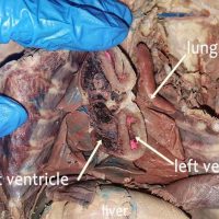
An “in situ” view is one that leaves the structure in it’s original place. In the view below, the heart of the cat has been cut to reveal the left and right ventricle. Note that the left ventricle has a much thicker muscular wall. This is because it …
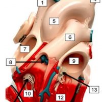
Heart models can be useful for learning the structure of the heart, particularly when paired with a dissection. They can be purchased at a reasonable cost at Amazon, usually mounted in a way that you can see the external and internal anatomy. When doing the dissection of …
The chordae tendinae are tiny tendons that connect the valves of the heart to the papillary muscle. They appear as small white strings and are sometimes referred to as the “heart strings.” In the photo below the right ventricle has been cut and the chordae shown are attached to …
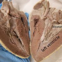
The bicuspid and tricuspid valves are both located between the atrium and the ventricle. They are similar in appearance and can be difficult for beginning anatomists to identify accurately. There are a couple of different ways to remember and then locate the valves. First, you must remember …
The heart and lungs of mammals are closely associated. You can find the lungs located on either side of the heart and often toward the dorsal side of the thoracic cavity. The lungs are soft, spongy tissue that are attached to the heart by the pulmonary artery and …
Often students mistake the pulmonary artery for the aorta. The pulmonary artery, or pulmonary trunk, is the most anterior vessel found on the heart; it is indicated below with a blue pencil. The aorta lies behind the pulmonary trunk, indicated with the red pencil. When the heart is …
This image of the heart shows a close-up view of the coronary vessels that are located on its surface. The cat has been injected with latex to color the vessels (blue for veins, pink for arteries.) Also visible is the pulmonary artery and the aorta.
The green pin in this heart indicates the pulmonary trunk which will split into the left and right pulmonary arteries. It is the most anterior vessel of the heart and carries blood from the heart to the lungs where blood becomes oxygenated and returns back to the …
The vessels of the heart are identified in this video: aorta, pulmonary trunk, brachiocephalic, and the vena cava. The heart is then cut in half the the internal structures are revealed: the atria, ventricles, bicuspid and tricuspid.
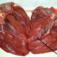
This dissected heart shows the left ventricle and the muscular wall of the septum. The left side of the heart is much more muscular than the right side of the heart, which is why we seem to feel our heartbeat more on the left side of the chest. …
Photo of the heart of a cat shows the major vessels: aorta, brachicephalic, common carotid, subclavian, and coronary vessels.