Tag: anatomy
The pineal gland (also called pineal body) is located in the midbrain and is one of the few non-paired structures of the brain. The pineal gland is part of the endocrine system and produces the hormone melatonin. Melatonin is associated with sleep patterns and circadian rhythms. …
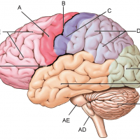
A = motor cortex | B = central sulcus | C = sensory cortex | D= parietal lobe | E = occipital lobe | AB = temporal lobe | AC = cerebellum | AD = medulla oblongata | AE = pons | BD = lateral fissure | BE = …
The bones of the ankle are collectively called the tarsals. The largest bone of this group is the calcaneus (9), or the heel bone. The talus (8) has an irregular shape and is commonly referred to as the “instep.” The navicular bone (7) is named for its …
a = epiphysis b = diaphysis c = articular cartilage d = periosteum f = compact bone g = medullary cavity (yellow marrow) h = endosteum j = epiphyseal line (growth plate) Coloring worksheet for this image.
This image of the heart shows a close-up view of the coronary vessels that are located on its surface. The cat has been injected with latex to color the vessels (blue for veins, pink for arteries.) Also visible is the pulmonary artery and the aorta.
A = retina | B = choroid | C = sclera | D = cornea E = lens | F = pupil | G = iris | H = suspensory ligaments I = optic disk | J = optic nerve | K = fovea centralis (macula) X …
This image shows a dissected cow eye. The vitreous humor has been removed and the retina is visible a thin layer of tissue covering the shiny blue tapetum. In cows, the tapetum is reflective and helps the animal see in the dark. (This is also why cow eyes …
The esophagus is simple to find on diagrams, usually shown as a long tube leading from the mouth to the stomach. On preserved specimens the esophagus can be challenging to locate. It is soft tissue that lies next to the trachea (identifiable by the cartilage rings) …