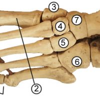
1. Phalanges (distal (a), middle (b), proximal (c) 2. Metatarsals 3. Medial Cuneiform 4. Intermediate Cuneiform 5. Medial Cuneiform 6. Cuboid 7. Navicular 8. Talus 9. Calcaneus
The bones of the ankle are collectively called the tarsals. The largest bone of this group is the calcaneus (9), or the heel bone. The talus (8) has an irregular shape and is commonly referred to as the “instep.” The navicular bone (7) is named for its …
This structure is names for its resemblance to a “turkish chair” and is found as a saddle-shaped depression on the sphenoid bone. The indentation (or the seat) is where the pituitary gland is located. To find the sella turcica, locate the sphenoid bone from the inside of the …
The top two vertebrae of the spine, the axis and the atlas form a joint (articulation) with the skull. The superior articular facet of the atlas, shown in the photo (blue dot ) articulates with the occipital condyle on the lower surface of the skull. The occipital condyles …
The cervical vertebrae are the first 7 vertebrae of the neck. The first two (C1 and C2) are the axis and the atlas and have unique features associated with their role of holding up the skull and providing a pivot joint around which the skull can rotate. …
The first two cervical vertebrae are the atlas (C1) and the axis (C2.) The atlas is named for the character from Greek Mythology who supported the globe, though in anatomy, the head is the globe. In fact, the articular facets of the atlas align perfectly with the occipital …
This is one of the hardest groups of bones to commit to memory and you will likely forget it if you don’t use it often. College professors and some high school anatomy teachers might ask you to learn the bones of the wrist. The task is made even …
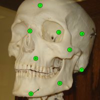
1. frontal 2. nasal 3. sphenoid 4. temporal 5. zygomatic 6. maxilla 7. mastoid process 8. mandible 9. mental foramen
A = cerebrum (parietal lobe) B = gyri (convolutions) C = corpus callosum D = frontal lobe E = thalamus F = hypothalamus G = pituitary gland H = midbrain J = pons K= medulla L = cerebellum (showing arbor vitae) M = transverse fissure N = …
a = epiphysis b = diaphysis c = articular cartilage d = periosteum f = compact bone g = medullary cavity (yellow marrow) h = endosteum j = epiphyseal line (growth plate) Coloring worksheet for this image.
This video shows the a sagittal cut in the brain of a sheep and identifies the major structures of the brain such as the cerebrum, corpus callosum, cerebellum, pons, medulla, thalamus, hypothalamus, pineal gland, and the four ventricles. https://youtu.be/Z9VFVFoOOo0
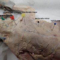
Dissected cat shows the main muscles of the thoracic region: the pectoantebrachialis, pectoralis major, pectoralis minor and the xiphihumeralis.