A = brachiocephalic veinB = mammary veinC = superior vena cavaD = pulmonary veinE = external jugular veinF = subclavian veinG = subclavian arteryH = brachiocephalic arteryI = aorta
The sheep brain shown below has the frontal and parietal lobe of the cerebrum pinned. The frontal lobe is responsible for cognitive functions such as learning and decision making. The parietal lobe is involved in how the brain processes and interprets sensory information. The cerebellum is pinned also, …
The liver is a large organ located at the top of the abdominal cavity and divided into several lobes. It is only found in vertebrates and it has several functions: detoxification, protein synthesis, and production of bile and other chemicals needed for digestion. Bile produced by the liver …
A = mesenteryB = large intestine (ascending colon)C = ileocecal valveD = cecumE = ileum of the small intestine
The brain stem of the sheep is located on the ventral surface of the brain. Visible features include the olfactory lobe, optic chiasma, pituitary stalk (infundibulum), midbrain, pons, medulla oblongata and the spinal cord. The image below shows the brain of the sheep with the dura mater removed.
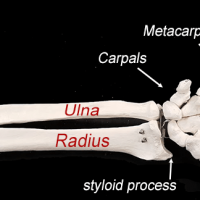
The radial notch is found on the ULNA and articulates with the head of the radius to form a pivot joint. It is located to the lateral side of the coronoid process. The coronoid and olecranon process articulate with the humerus to form the hinge joint of the …
Learning the names of the tarsals can be challenging. If you can locate the navicular bone, which articulates with talus, then the cuneiform bones are easy to locate. Navicular means “boat”, so just think of the three cuniform bones as people on the boat. The one closest to …
An illustration of a male and female pelvis showing the differences between the shape of the ileum, the angle between the right and left ischium (pubic arch). The sacrum in females is also much wider. These features create a larger opening in the pelvis, an adaptation for childbirth.
The placenta is connected to the umbilical cord and attaches to the wall of the uterus. This organ allows for nutrient exchange and the removal of wastes. The placenta develops when the blastocyst (embryo) implants into the uterine wall. Usually this attachment occurs at the superior end of …
Capsule Cortex Renal Pyramids Renal Tubules Malpighian corpuscles Renal Papillae Medulla Renal Pelvis Ureter Renal Vein Renal Artery Hilus Calyx Interlobar arteries and veins
Left gastric vein (coronary vein) Spleen Splenic vein Gastroepiploic vein Inferior mesenteric vein Left colic vein Jejunal vein Sigmoid vein Hemorrhoidal vein Ileocolic vein Right colic vein Median colic vein Superior mesenteric vein Hepatic portal vein Inferior vena cavaa Right hepatic vein Liver
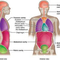
Most anatomy textbooks begin with descriptions of body cavities and body regions. These terms are important for identifying the location of structures. For example, you will fund the lungs within the thoracic cavity, which is the region superior to the diaphragm. The stomach and digestive organs are …