1. Posterior Commissure 2. Anterior Commissure 3. Lamina of septum pelucidum 4. Genu of corpus callosum 5. Fornix 7. Thalamus 8. Choroid plexus 9. Splenium of corpus callosum 10. Great vein of cerebrum 11. Pineal Body 12. Quadrigeminal body (superior and inferior colliculi) 13. Cerebellum 14. Medulla …
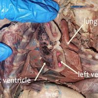
An “in situ” view is one that leaves the structure in it’s original place. In the view below, the heart of the cat has been cut to reveal the left and right ventricle. Note that the left ventricle has a much thicker muscular wall. This is because it …
To view the internal structures of the kidney, a coronal section can be applied. This type of cut will separate the front half of the structure from the back half. The inner area is called the medulla and is made of individual renal pyramids, which may not …
The spleen is involved in the immune system and is responsible for removing old blood cells. It sits under the rib cage in humans on the left side of the abdomen. In the cat, the spleen is an elongated structure that is often reddish-brown in color on preserved …
This x-ray was taken by a dentist, showing two wisdom teeth (white arrows) positioned below the gum line. Most people have three permanent sets of molars: the first set usually appears at around the age of 6, and then the 2nd set appears at around the age …
In February of 2016, model Katie May died suddenly after a visit to a chiropractor. The official cause of death was “vertebral artery dissection.” During spinal manipulation, the artery which supplies blood to brain was damaged. The brain’s arteries circle the brain around the pituitary gland consisting …
The esophageal hiatus is the opening in the diaphragm through which the esophagus passes and then enters the stomach. It can be viewed by pushing aside the lungs and heart within the thoracic cavity and tracing the esophagus to the diaphragm. The inferior vena cava also can …
Answers: A= Renal Vein B= Right Kidney C= Inferior Vena Cava D = Urinary Bladder E= Renal Artery AB = Right Kidney AC = Abdominal Aorta AD = Ureter AE = Urethra
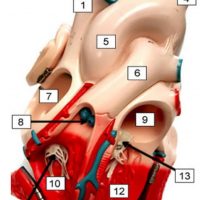
Heart models can be useful for learning the structure of the heart, particularly when paired with a dissection. They can be purchased at a reasonable cost at Amazon, usually mounted in a way that you can see the external and internal anatomy. When doing the dissection of …
The small intestine connects to the colon where a valve regulates the movement of food. The ileocecal valve is named for the ileum and the cecum it connects to. The cecum is a pouch-like section of the large intestine that is the first part of the colon, also called …
The gall bladder is a structure found under the liver of many vertebrates, including humans, where it stores bile that is secreted by the liver. Bile is transported to the duodenum through the bile duct; its main function is to aid in the digestion of fats. In humans, …
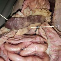
The pancreas can be one of the more difficult structures for beginning anatomy students. This is partly because it is a gland that doesn’t preserve well and can fall apart easily when handling the stomach or other organs. In ideal specimens, the pancreas can be seen as a bumpy …