Tag: renal
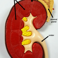
Capsule Cortex Renal Pyramids Renal Tubules Malpighian corpuscles Renal Papillae Medulla Renal Pelvis Ureter Renal Vein Renal Artery Hilus Calyx Interlobar arteries and veins
To view the internal structures of the kidney, a coronal section can be applied. This type of cut will separate the front half of the structure from the back half. The inner area is called the medulla and is made of individual renal pyramids, which may not …
Answers: A= Renal Vein B= Right Kidney C= Inferior Vena Cava D = Urinary Bladder E= Renal Artery AB = Right Kidney AC = Abdominal Aorta AD = Ureter AE = Urethra
Renal pyramids are triangular shaped areas seen on a cross section of the kidney. They appear striped due to the thousands of nephrons within them that make up the functional unit of the kidney. The nephrons perform the function of filtration of waste products from the blood and …
A = interlobar arteries and veins B = renal artery C = renal vein D = ureter E = renal pyramids F = minor calyx G = major calyx H = renal pelvis I = capsule J = medulla K = cortex L = nephrons
The abdominal aorta is the largest artery of the abdominal cavity. The aorta originates at the heart, forms and arch and then continues as the descending aorta. Above the diaphragm, it is called the thoracic aorta; below the diaphragm, it is called the abdominal aorta. There are …
This image shows the inferior vena cava where it branches to the renal vein leading to the kidney. The vessels appear blue because the cat has been injected with blue latex.
This video traces the abdominal aorta to show where it branches to the celiac trunk, the superior mesenteric, renal arteries and inferior mesenteric. The inferior vena cava is also shown with the renal vein branches. Finally, the abdominal aorta splits to travel into the legs at the …