Tag: dissection
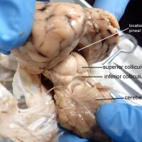
The brain stem of the sheep is located on the ventral surface of the brain. Visible features include the olfactory lobe, optic chiasma, pituitary stalk (infundibulum), midbrain, pons, medulla oblongata and the spinal cord. The image below shows the brain of the sheep with the dura mater removed.
Students in biology are often tasked with dissecting a frog, but there are many other vertebrate models that can provide a rich experience in learning anatomy. Common specimens include the fetal pig, shark, mink, cat, and the rat. Rats are fairly inexpensive and are raised for …
This video shows the major parts of the brain, starting with the external anatomy where the pituitary gland, optic chiasma (nerves) , olfactory lobes, dura mater, and the parts of the brain stem: midbrain, pons, medulla, and spinal cord. The brain is the cut along the longitudinal …
The superior colliculus is part of the midbrain, also known as the tectum in non-mammalian vertebrates. This area of the brain directs behavioral responses, particularly how the eyes respond to stimulus and the corresponding movement of the head and neck. If you are looking for this structure in …
Often students mistake the pulmonary artery for the aorta. The pulmonary artery, or pulmonary trunk, is the most anterior vessel found on the heart; it is indicated below with a blue pencil. The aorta lies behind the pulmonary trunk, indicated with the red pencil. When the heart is …
The urinary bladder collects urine excreted by the kidneys. It is a hollow, expandable structure located in the lower abdomen. The photo below shows the urinary bladder of a sheep. The bladder is the balloon-like structure in the upper right area of the tray. It is …
Procedural video showing how to dissect the eye and identify the major structures such as the lens, iris, pupil, vitreous humor, retina, optic disk, optic nerve. and tapetum. Unlike humans, cows have a reflective surface on the back of their eye which enables them to see in the …
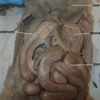
This image shows the stomach and the first section of the small intestine, called the duodenum. The next section of the small intestine is the jejunum followed by the ileum which connects it to the large intestine.
This image shows a dissected cow eye. The vitreous humor has been removed and the retina is visible a thin layer of tissue covering the shiny blue tapetum. In cows, the tapetum is reflective and helps the animal see in the dark. (This is also why cow eyes …
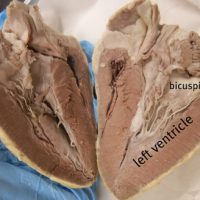
The vessels of the heart are identified in this video: aorta, pulmonary trunk, brachiocephalic, and the vena cava. The heart is then cut in half the the internal structures are revealed: the atria, ventricles, bicuspid and tricuspid.
The esophagus is simple to find on diagrams, usually shown as a long tube leading from the mouth to the stomach. On preserved specimens the esophagus can be challenging to locate. It is soft tissue that lies next to the trachea (identifiable by the cartilage rings) …
Finding the superior and inferior mesenteric arteries takes patience. Carefully tease away the connective tissue around the large intestine and find where it attaches to the abdominal aorta. A = abdominal aorta B = celiac trunk C = superior mesenteric artery D = inferior mesenteric artery