Tag: aorta
A = brachiocephalic veinB = mammary veinC = superior vena cavaD = pulmonary veinE = external jugular veinF = subclavian veinG = subclavian arteryH = brachiocephalic arteryI = aorta
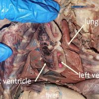
An “in situ” view is one that leaves the structure in it’s original place. In the view below, the heart of the cat has been cut to reveal the left and right ventricle. Note that the left ventricle has a much thicker muscular wall. This is because it …
Heart models can be useful for learning the structure of the heart, particularly when paired with a dissection. They can be purchased at a reasonable cost at Amazon, usually mounted in a way that you can see the external and internal anatomy. When doing the dissection of …
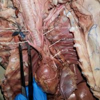
The carotid arteries supply blood to the head and neck. In humans, the right common carotid branches from the brachiocephalic artery and the left common carotid branches directly from the arch of the aorta. On the cat, as shown in the photograph, both common carotids branch from the …
During the cardiac cycle, two contractions occur. Systole occurs when the muscles contract and diastole occurs when the muscles relax. Atrial systole occurs when the atria contract and push blood into the lower ventricles. During this point, the aortic valve is closed. Ventricular contraction then occurs …
Identify the vessels. 1. Inferior Vena Cava 2. Abdominal Aorta 3. Iliolumbar artery 4. External Iliac 5. Internal Iliac
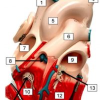
Often students mistake the pulmonary artery for the aorta. The pulmonary artery, or pulmonary trunk, is the most anterior vessel found on the heart; it is indicated below with a blue pencil. The aorta lies behind the pulmonary trunk, indicated with the red pencil. When the heart is …
The celiac trunk (artery) is the first major branch of the abdominal aorta, as shown in the photo below. The celiac artery then branches into the gastric artery, which goes to the stomach, the hepatic artery that goes to the liver, and the splenic artery that goes to …
On the cat, the external iliac arteries bifurcate off the abdominal aorta, forming a Y. The internal iliac arteries also form a Y on the inside of the external iliac arteries. The external iliac arteries will continue into the hind limbs of the cat and become …
The abdominal aorta is the largest artery of the abdominal cavity. The aorta originates at the heart, forms and arch and then continues as the descending aorta. Above the diaphragm, it is called the thoracic aorta; below the diaphragm, it is called the abdominal aorta. There are …
This image of the heart shows a close-up view of the coronary vessels that are located on its surface. The cat has been injected with latex to color the vessels (blue for veins, pink for arteries.) Also visible is the pulmonary artery and the aorta.
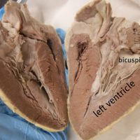
The vessels of the heart are identified in this video: aorta, pulmonary trunk, brachiocephalic, and the vena cava. The heart is then cut in half the the internal structures are revealed: the atria, ventricles, bicuspid and tricuspid.
This video traces the abdominal aorta to show where it branches to the celiac trunk, the superior mesenteric, renal arteries and inferior mesenteric. The inferior vena cava is also shown with the renal vein branches. Finally, the abdominal aorta splits to travel into the legs at the …
Finding the superior and inferior mesenteric arteries takes patience. Carefully tease away the connective tissue around the large intestine and find where it attaches to the abdominal aorta. A = abdominal aorta B = celiac trunk C = superior mesenteric artery D = inferior mesenteric artery
Photo of the heart of a cat shows the major vessels: aorta, brachicephalic, common carotid, subclavian, and coronary vessels.
If you trace the aorta into the abdomen, it will eventually branch into two large vessels that go into the lower extremities, the external iliac arteries. Smaller vessels, the internal iliac vessels supply blood to the genitals.
This video shows the heart and the major vessels that branch from it on a dissected cat. The superior vena cava, aorta, subclavian, and brachiocephalic vessels are shown clearly to help students identify them on their own specimens.