Author: anatomycorner
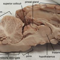
The brain stem is the posterior section of the brain that includes the pons and the medulla oblongata, which is continuous with the spinal cord. The midbrain is usually included as part of the brain stem and includes the corpora quadrigemina (shown partially on the photo as the …
On the ventral side of the sheep’s brain, several nerves and structures are visible after the careful removal of the dura mater. The image below shows the brain with the dura intact and the eye socket attached. The blue reflective surface of the sheep tapetum can be …
In the cat, the trapezius muscle is divided into three flat muscles that cover the upper back and the neck. The most anterior of these muscles is the clavotrapezius, named for its insertion into the clavicle. The middle trapezius muscle is the acromiotrapezius, which covers the scapula and …
This bisected brain shows the major features of the midbrain and brain stem. The corpus callosum is just been the cortex of the brain and connects the left and right hemispheres. The lateral ventricle is a fluid filled space found below the corpus callosum and just about …
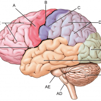
The cerebrum and cerebellum of the brain are divided by the transverse fissure. The left and right hemispheres of the brain are divided by the longitudinal fissure. A fissure is a groove or a natural division, and with the brain are divide major regions. Sulci (singular: sulcus) are …
The pineal gland (also called pineal body) is located in the midbrain and is one of the few non-paired structures of the brain. The pineal gland is part of the endocrine system and produces the hormone melatonin. Melatonin is associated with sleep patterns and circadian rhythms. …
A = motor cortex | B = central sulcus | C = sensory cortex | D= parietal lobe | E = occipital lobe | AB = temporal lobe | AC = cerebellum | AD = medulla oblongata | AE = pons | BD = lateral fissure | BE = …
Practice naming the neuroglial cells.
Neuroglial cells are sometimes simply called “glia” which in Greek, means “glue.” These cells support the neurons by providing insulation, supplying nutrients, and removing dangerous pathogens. Each cell of the glia can be identified on a diagram by examining the general location, attachment to other stuctures and …
The sartorius muscle is the longest muscle in the human body, extending from the hip to the inside of the thigh. On the cat, this muscle is much more compact, and covers the thigh and knee joint as a superficial muscle. It is shown on the photo …
This cat illustrates the three pectoralis muscles: the pectoantebrachialis, the pectoralis major, and the pectoralis minor. One way to find the pectoantebrachialis is to stretch the forelimbs of the cat which will reveal the bordering fascia. This muscles goes straight across the chest and to the forelimbs. …
The zygomatic bone articulates with the maxilla and the temporal bone and is the bone that makes up the cheek of the face. The zygomatic process of the temporal bone is an extension of the temporal bone that connects to the zygomatic bone. On the …
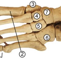
1. Phalanges (distal (a), middle (b), proximal (c) 2. Metatarsals 3. Medial Cuneiform 4. Intermediate Cuneiform 5. Medial Cuneiform 6. Cuboid 7. Navicular 8. Talus 9. Calcaneus
The bones of the ankle are collectively called the tarsals. The largest bone of this group is the calcaneus (9), or the heel bone. The talus (8) has an irregular shape and is commonly referred to as the “instep.” The navicular bone (7) is named for its …
This structure is names for its resemblance to a “turkish chair” and is found as a saddle-shaped depression on the sphenoid bone. The indentation (or the seat) is where the pituitary gland is located. To find the sella turcica, locate the sphenoid bone from the inside of the …
The top two vertebrae of the spine, the axis and the atlas form a joint (articulation) with the skull. The superior articular facet of the atlas, shown in the photo (blue dot ) articulates with the occipital condyle on the lower surface of the skull. The occipital condyles …
The cervical vertebrae are the first 7 vertebrae of the neck. The first two (C1 and C2) are the axis and the atlas and have unique features associated with their role of holding up the skull and providing a pivot joint around which the skull can rotate. …
The first two cervical vertebrae are the atlas (C1) and the axis (C2.) The atlas is named for the character from Greek Mythology who supported the globe, though in anatomy, the head is the globe. In fact, the articular facets of the atlas align perfectly with the occipital …