Author: anatomycorner
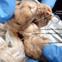
The cerebellum is located at the posterior end of the brain and is associated with motor control, balance and coordination. It is only loosely attached to the cerebrum, and dissections of the brain reveal that removing the dura mater often result in the accidental removal of the cerebellum. …
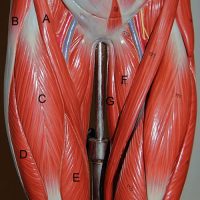
The thigh muscles are often collectively called the quadriceps, which consist of four separate muscles: rectus femoris, vastus lateralis, vastus medialis, and the vastus intermedius. On most diagrams, the rectus femoris is shown as the front muscle that runs parallel to the femur. The word “rectus” means …
The lower arm consists of two bones, the radius and the ulna. The ulna articulates with the humerus to form a hinge joint. The radius articulates with the ulna at the radial notch and forms a pivot joint, allowing for the rotation of the wrist. This type of …
The ulna is one of two bones found in the forearm. It forms a hinge joint with the humerus and a pivot joint with the radius, giving the lower arm the range of motion to bend and rotate the hands. On anatomical drawings, the ulna and radius …
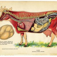
You may have heard that cows have 4 stomachs, but that is not actually accurate. Cows, like all other vertebrates, have the same basic blueprint for the digestive system: mouth –> esophagus –> stomach –> small intestine –> large intestine –> anus There is one stomach, but that stomach is …
The superior colliculus is part of the midbrain, also known as the tectum in non-mammalian vertebrates. This area of the brain directs behavioral responses, particularly how the eyes respond to stimulus and the corresponding movement of the head and neck. If you are looking for this structure in …
Epithelial tissue serves as a covering for external structures (skin) and internal organs. There are many categories of epithelial cells that many beginning anatomy students must learn to identify. Generally, epithelial tissue is classified as either simple or stratified. Stratified tissue consists of many layers of cells …
The epiglottis is elastic cartilage that is attached to the entrance of the larynx and prevents food and liquids from entering the airway. In humans, the larynx is positioned much lower in body, which results in an increased risk of choking. The position of the voice box also …
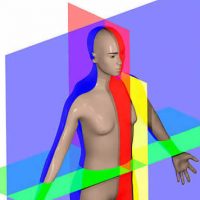
The planes of the body are one of the first topics in Anatomy and Physiology. The concept relies on understanding the orientation of the specimen. Sagittal = cuts the body into a left and right side (red) Coronal = cuts the body into a front and back …
Osteocytes are bone cells that sit within chambers called lacuna. Lacuna are connective via tine canals called canaliculi and form concentric circles around a central haversian canal. This coloring worksheet may help with your understanding of these relationships. It can be very difficult to develop a …
The clavicle is commonly called the collarbone and it located in the upper chest region. The word “clavicle” is latin for “little key” because it rotates like a key when the shoulder and arm are extended. The clavicle connects to the acromion process of the scapula and the …
cranial dorsal thoracic spinal ventral diaphragm (muscle) abdominal abdominopelvic pelvic
The lower leg bones of the human consist of the large tibia, which is commonly called the shinbone, and the smaller fibula which is located laterally. Several features of these bones are visible from the outside of the body. The large tibial tuberosity, is visible as a bulge just …
The lower end of the femur, called the distal end, articulates with the tibia of the lower leg. It consists of two large structures called condyles, which is the greek word for “knuckle.” In fact, if you go to a butcher shop, you can order a …
The proximal end of the femur articulates with the hip. The rounded head of the femur fits within the acetabulum of the coxal bones, forming a ball-and-socket joint. The fovea capitis is a small depression in the head serves as an attachment point for ligaments. The greater trochanter …
Identify the vessels. 1. Inferior Vena Cava 2. Abdominal Aorta 3. Iliolumbar artery 4. External Iliac 5. Internal Iliac
The iliolumbar arteries are named for their location, supplying blood to the lumbar region of the back and the ilium of the of the pelvis. They branch from the abdominal aorta just above where it splits into the external and internal iliac arteries.
The trachea is also called the windpipe and it connects the mouth to the lungs. It is distinguished from the nearby esophagus by the presence of cartilage rings which prevent the airway from collapsing. At the top of the trachea is the larynx, or the voicebox. …