
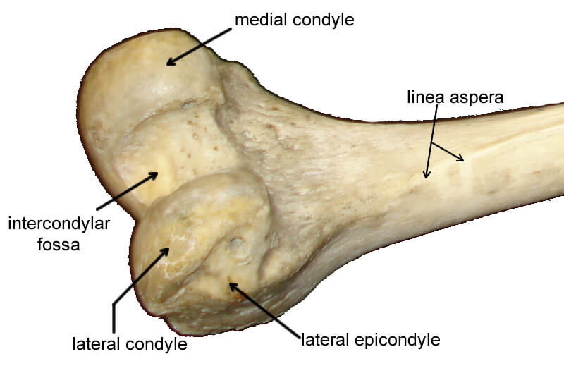
 The lower end of the femur, called the distal end, articulates with the tibia of the lower leg. It consists of two large structures called condyles, which is the greek word for “knuckle.” In fact, if you go to a butcher shop, you can order a “knuckle bone” for soups or for dog treats. The two condyles are named medial condyle and lateral condyle based on their positioning.
It can be challenging to determine the medial and lateral side on a disarticulated bone. The best way is to look a the top of the bone where the “knob-like” head of the femur is, the head points toward the body, this is the medial side.
Each condyle has a smaller protuberance called the epicondyles, and the deep groove between the condyles is called the intercondylar fossa. Also visible on the femur is the linea aspera (Latin = “rough line”) which serves as an attachment point for muscles of the thigh.


