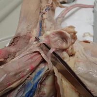
This image shows the superior vena cava and the two brachiocephalic veins that branch from it. The brachiocephalic vein then branches into the subclavian artery and the external jugular veins.
The urinary bladder is found in the lower abdominal cavity, shown here with the two ureters that drain urine from the kidneys into the bladder.
This video shows the heart and the major vessels that branch from it on a dissected cat. The superior vena cava, aorta, subclavian, and brachiocephalic vessels are shown clearly to help students identify them on their own specimens.
A. Esophagus B. Cardiac Sphincter Valve C. Fundus D. Cardiac region of the stomach E. Body of the stomach AB. Rugae AC. Pyloric Region of the stomach AD. Pyloric Sphincter Valve AE. Duodenum of …
The ureters are tubes that connect the kidney to the urinary bladder. The tubes are thin and sometimes difficult to find. One way to locate the ureter on a specimen is to wiggle the kidney, which will then reveal the location of the ureter. The ureter and …
I was finally was able to compile and organize all the photos taken of the cat dissection. The virtual cat dissection is simply a collection of pages that walk you through the dissection and point out various structures. Photos are labeled to help supplement a real dissection done …
Anatomy classes of 2010-2011 dissected a cat. Throughout the year, I took photos of the dissection. I have begun the process of cleaning up the photos, deleting and categorizing so that I can improve on the galleries of this site. Right now, the galleries are categorized by …
Starting to put together this website, mainly following the same format as biologycorner.com. I did want to use new colors though to give this site a distinctive feel. Biologycorner only has limited human anatomy resources, buried in the classes pages. This site will be more general to serve …