Category: Anatomy
Finding the superior and inferior mesenteric arteries takes patience. Carefully tease away the connective tissue around the large intestine and find where it attaches to the abdominal aorta. A = abdominal aorta B = celiac trunk C = superior mesenteric artery D = inferior mesenteric artery
Yellow = gastrocnemius | Red = biceps femoris | Blue = semitendinosis
Photo of the heart of a cat shows the major vessels: aorta, brachicephalic, common carotid, subclavian, and coronary vessels.
If you trace the aorta into the abdomen, it will eventually branch into two large vessels that go into the lower extremities, the external iliac arteries. Smaller vessels, the internal iliac vessels supply blood to the genitals.
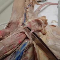
This image shows the superior vena cava and the two brachiocephalic veins that branch from it. The brachiocephalic vein then branches into the subclavian artery and the external jugular veins.
The urinary bladder is found in the lower abdominal cavity, shown here with the two ureters that drain urine from the kidneys into the bladder.
The ureters are tubes that connect the kidney to the urinary bladder. The tubes are thin and sometimes difficult to find. One way to locate the ureter on a specimen is to wiggle the kidney, which will then reveal the location of the ureter. The ureter and …