Author: anatomycorner
This video traces the abdominal aorta to show where it branches to the celiac trunk, the superior mesenteric, renal arteries and inferior mesenteric. The inferior vena cava is also shown with the renal vein branches. Finally, the abdominal aorta splits to travel into the legs at the …
The pectoralis muscles are divided into three sections: pectoantebrachialis, pectoralis major, pectoralis minor. The xiphihumeralis is named for its connection between the humerus and the xiphoid process of the sternum.
This dissected heart shows the left ventricle and the muscular wall of the septum. The left side of the heart is much more muscular than the right side of the heart, which is why we seem to feel our heartbeat more on the left side of the chest. …
The esophagus is simple to find on diagrams, usually shown as a long tube leading from the mouth to the stomach. On preserved specimens the esophagus can be challenging to locate. It is soft tissue that lies next to the trachea (identifiable by the cartilage rings) …
These brains are shipped with the dura mater intact. Students carefully remove the dura to expose the soft tissue of the cerebrum underneath.
Finding the superior and inferior mesenteric arteries takes patience. Carefully tease away the connective tissue around the large intestine and find where it attaches to the abdominal aorta. A = abdominal aorta B = celiac trunk C = superior mesenteric artery D = inferior mesenteric artery
Yellow = gastrocnemius | Red = biceps femoris | Blue = semitendinosis
Photo of the heart of a cat shows the major vessels: aorta, brachicephalic, common carotid, subclavian, and coronary vessels.
Answers: A. Parotid Salivary Gland B. Tongue C. Sublingual Salivary Gland D. Esophagus E. Cardiac Sphincter Valve AB. Liver AC. Gall Bladder AD. Ascending Colon AE. Cecum …
A. Esophagus B. Cardiac Sphincter Valve C. Stomach D. Pancreas E. Duodenum AB. Gall Bladder AC. Liver
A. Stomach B. Duodenum C. Jejunum (small intestine) D. Ascending Colon AB Cecum AC. Mesentery AD. Appendix AE. Ileum (small intestine)
If you trace the aorta into the abdomen, it will eventually branch into two large vessels that go into the lower extremities, the external iliac arteries. Smaller vessels, the internal iliac vessels supply blood to the genitals.
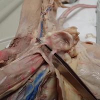
This image shows the superior vena cava and the two brachiocephalic veins that branch from it. The brachiocephalic vein then branches into the subclavian artery and the external jugular veins.
The urinary bladder is found in the lower abdominal cavity, shown here with the two ureters that drain urine from the kidneys into the bladder.
This video shows the heart and the major vessels that branch from it on a dissected cat. The superior vena cava, aorta, subclavian, and brachiocephalic vessels are shown clearly to help students identify them on their own specimens.
A. Esophagus B. Cardiac Sphincter Valve C. Fundus D. Cardiac region of the stomach E. Body of the stomach AB. Rugae AC. Pyloric Region of the stomach AD. Pyloric Sphincter Valve AE. Duodenum of …
The ureters are tubes that connect the kidney to the urinary bladder. The tubes are thin and sometimes difficult to find. One way to locate the ureter on a specimen is to wiggle the kidney, which will then reveal the location of the ureter. The ureter and …
I was finally was able to compile and organize all the photos taken of the cat dissection. The virtual cat dissection is simply a collection of pages that walk you through the dissection and point out various structures. Photos are labeled to help supplement a real dissection done …