Category: Video
This video shows the major parts of the brain, starting with the external anatomy where the pituitary gland, optic chiasma (nerves) , olfactory lobes, dura mater, and the parts of the brain stem: midbrain, pons, medulla, and spinal cord. The brain is the cut along the longitudinal …
Procedural video showing how to dissect the eye and identify the major structures such as the lens, iris, pupil, vitreous humor, retina, optic disk, optic nerve. and tapetum. Unlike humans, cows have a reflective surface on the back of their eye which enables them to see in the …
This video shows the a sagittal cut in the brain of a sheep and identifies the major structures of the brain such as the cerebrum, corpus callosum, cerebellum, pons, medulla, thalamus, hypothalamus, pineal gland, and the four ventricles.
Video shows the major structures: cerebrum, transverse fissure, longitudinal fissure, superior and inferior colliculi, pineal gland, cerebellum.
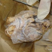
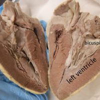
The vessels of the heart are identified in this video: aorta, pulmonary trunk, brachiocephalic, and the vena cava. The heart is then cut in half the the internal structures are revealed: the atria, ventricles, bicuspid and tricuspid.
This video traces the abdominal aorta to show where it branches to the celiac trunk, the superior mesenteric, renal arteries and inferior mesenteric. The inferior vena cava is also shown with the renal vein branches. Finally, the abdominal aorta splits to travel into the legs at the …
This video shows the heart and the major vessels that branch from it on a dissected cat. The superior vena cava, aorta, subclavian, and brachiocephalic vessels are shown clearly to help students identify them on their own specimens.